
08 Jun 2023
Going Viral: Exploring Viruses in Virtual Reality
Led by Dr Jason Roberts and Jamie Mumford of the Doherty Institute's Electron Microscopy and Structural Virology Laboratory, students in the University of Melbourne’s Master of Biomedical Science recently embarked on a journey of scientific exploration that merged technology, public health and academia like never before.
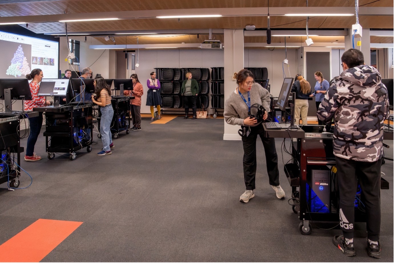
Students get ready to don headsets and explore viruses in virtual reality
The Royal Melbourne Hospital’s Dr Jason Roberts, Head of the Electron Microscopy and Structural Virology Laboratory at Doherty Institute, demonstrated to students how a modern virologist works in virtual reality (VR), using samples from his own published work.
Doning VR headsets, the students immersed themselves inside an infectious virus particle. This new cutting-edge learning experience lets students visualise real-world applications of VR as used in the intersecting disciplines of public health and basic research.
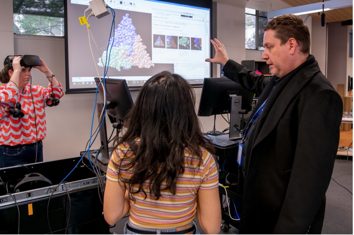
Dr Jason Roberts (right) teaches students how to manipulate virus structures in virtual reality.
Under Dr Roberts' expert guidance, the students delved into the intricate world of viruses by examining the three-dimensional structure of the insect virus black beetle nodavirus in VR using ChimeraX, a next-generation molecular visualisation program. They learned to navigate through three-dimensional structures and to manipulate various display settings to gain a comprehensive understanding of the viral anatomy. Additionally, the students practiced aligning cryo-electron micrographs with atomic models, a crucial aspect of advanced virological research.
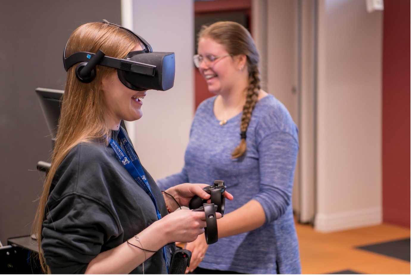
Students enjoyed using VR to learn about viruses.
Once the students had familiarised themselves with the intricacies of working with viruses in a virtual environment, they turned their attention to a sample of Mpox-infected cellular material provided by Dr Roberts[1]. Unlike traditional 2-dimensional screens, the VR technology allowed the students to freely explore the entire slice of infected cells and observe the distinctive dumbbell-like structures within the Mpox virions, that serve as a diagnostic hallmark of poxvirus infection.
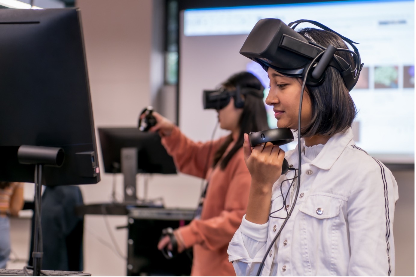
A student considers the next step in the workshop.
The use of VR in the subject Defence & Disease: Frontier Technologies not only provided a realistic and engaging experience, but also offered the students a unique perspective on the subject matter. By transcending the limitations of conventional learning methods, this immersive VR adventure granted the students a sense of freedom to examine infectious diseases in a more comprehensive and interactive manner, using the same technologies that experts in the field use daily.
Note: This article was adapted from an article originally posted on the website of the University of Melbourne’s School of Biomedical Sciences.
[1] Lim, C. K., Roberts, J., Moso, M., Liew, K. C., Taouk, M. L., Williams, E., Tran, T., Steinig, E., Caly, L., & Williamson, D. A. (2023). Mpox diagnostics: Review of current and emerging technologies. Journal of Medical Virology, 95(1). https://doi.org/10.1002/jmv.28429