-
Research Groups
-
Mueller Group
Research in Scott’s group is focused on examining immune responses to both acute and chronic viral infections. A particular emphasis on T cell responses and interactions with antigen presenting cells and lymphoid tissue stromal cells is currently driving the group, as well as an interest in neuro-immune interactions.
Other work areas include:Malaria
Current Projects
-
Priming of T Cell responses to viral infection
Much of our understanding of the complex series of events that occur during the priming of immune responses to infections or following vaccination have been gained via static methods such as flow cytometry, histology and cell culture. These methods provide limited information on when, where and how different cells interact during the different phases of immunity. Scott’s group now has the ability to follow immune cells within living tissues and animals to determine their behaviour in real time using intravital 2-photon microscopy. A major goal is to understand how T cells are activated in response to viral infections. T cells (both CD4+ and CD8+) are first activated in draining lymph nodes via interactions with dendritic cells (DC) presenting viral antigens. Using well-established models of localised or systemic viral infection, they are directly examining T cell responses by 2-photon microscopy in the skin, lymph nodes and spleen. They have assembled an array of powerful tools, including recombinant viruses and transgenic mice expressing fluorescent molecules in various compartments.Much of our understanding of the complex series of events that occur during the priming of immune responses to infections or following vaccination have been gained via static methods such as flow cytometry, histology and cell culture. These methods provide limited information on when, where and how different cells interact during the different phases of immunity. Scott’s group now has the ability to follow immune cells within living tissues and animals to determine their behaviour in real time using intravital 2-photon microscopy. A major goal is to understand how T cells are activated in response to viral infections. T cells (both CD4+ and CD8+) are first activated in draining lymph nodes via interactions with dendritic cells (DC) presenting viral antigens. Using well-established models of localised or systemic viral infection, they are directly examining T cell responses by 2-photon microscopy in the skin, lymph nodes and spleen. They have assembled an array of powerful tools, including recombinant viruses and transgenic mice expressing fluorescent molecules in various compartments.
-
Tissue-resident memory T cells in the skin
Protection from viral infections that target peripheral tissues such as the skin requires coordinated recruitment of pathogen-specific T lymphocytes into the tissue where they function to defeat the virus. Following the resolution of HSV-1 infection or inflammation in the skin, tissue-resident memory (Trm) CD8+ T cells reside for long periods in the epidermis where they can provide enhanced protection from reinfection. These tissue-resident T cells display a unique dendritic morphology and a slow pattern of tissue surveillance. Scott’s group is examining how these Trm cells are formed, persist and respond following secondary infection.
-
Nervous regulation of T cell migration and anti-viral immunity
The lymphoid organs are innervated by fibres of the sympathetic nervous system (SNS), which release SNS neurotransmitters during times of chronic stress. SNS neurotransmitters bind to beta-adrenoceptors (βARs) on multiple cell types to induce genomic and functional changes. However, little is known about the impact of SNS neurotransmitters on immune cell function or their role in anti-viral immunity. Studies have shown that viral immunity is compromised during times of chronic stress raising the possibility that SNS signalling impairs anti-viral immunity by impacting T cell activation, migration or effector functions. This project is examining the effect of adrenergic receptor signalling in T cells on their migration within tissues such as the lymph nodes and subsequent immune responses to viral infections.
-
Lymphoid stromal cell responses to infection
The lymphoid tissues (spleen and lymph nodes) are constructed of a complex microarchitecture that is supported by networks of endothelial and mesenchymal stromal cells. The T cell zones in the spleen and lymph nodes are supported by a network of stromal cells known as fibroblastic reticular cells (FRC). Little is known of the phenotype of these stromal cell subsets and how they can influence the immune cells they regularly interact with. Scott’s group is examining how FRC contribute to the expansion and contraction of secondary lymphoid organs during immune responses to acute and chronic viral infections, as well as following recall infection. The phenotypic changes that occur in the secondary lymphoid organ stromal cells after infection will reveal how FRC function changes and whether pathogens can differentially influence the functions of these critical cells and how this influences immunity.
-
Neural regulation of anti-cancer immunity
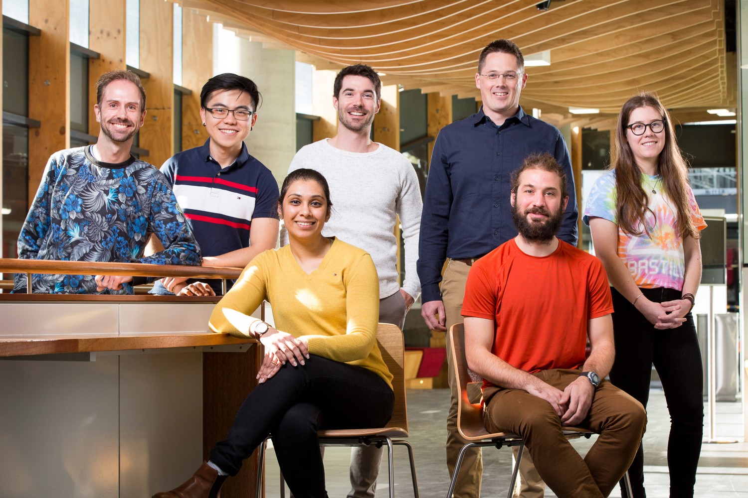
Lab Team
-
Dr Yannick AlexandrePost-Doc
-
Research Officer
-
Keit LoiPhD student
-
Sarah SandfordPhD student
-
Thomas HueneburgMasters student
-
Natasha ZamudioResearch Officer
-
Melanie DamtsisResearch Assistant
-
Barrow group
My lab is interested in how receptors expressed by immune cells distinguish normal healthy cells from malignant cells or pathogens. We want to understand how these receptors work together to regulate immune responses so we can design better clinical interventions.
-
Bedoui Group
The Bedoui Lab uses models of viral and bacterial infection to study how the innate and the adaptive immune system interact. Key foci are to understand how innate cells sense pathogens and how this information is integrated into protective adaptive T cell responses.
Other work areas include:Bacterial and Parasitic Infections
-
Brooks Group
Research in Andrew’s laboratory is largely centred on how natural killer cells and T cells impact the outcomes of viral infection, cancer and transplantation.
-
Chung group
The Chung group is interested in understanding the biophysical and functional properties of antibodies that are associated with protection against a range of infectious diseases, which will provide important insights to improve antibody-based vaccines and therapies.
Other work areas include:COVID-19, Viral Infectious Diseases
-
Corbett Group
Our team study mucosal-associated invariant T (MAIT) cells. We seek to understand how vitamin-based antigens are produced by microorganisms, how MAIT cells detect these antigens and how we can manipulate MAIT cell functions to improve outcomes in infectious and non-infectious diseases.
Other work areas include:Bacterial and Parasitic Infections
-
Gebhardt Group
Thomas’ group studies basic and translational aspects of immune responses in peripheral tissues. Their overall goal is to develop future vaccines and immunomodulatory therapies that target peripheral T cells for improved clinical outcomes in infection, inflammation and cancer.
-
Heath Group
Bill’s group’s cellular immunology research currently focuses on understanding killer T cell function with particular reference to improved vaccination strategies and understanding malarial disease.
Other work areas include:Malaria
-
Kallies Group
The Kallies group studies the molecular control of lymphocyte differentiation in response to antigen with a particular focus on T cells residing in non-lymphoid tissues, including tumors.
Other work areas include:Viral Infectious Diseases
-
Kedzierska Group
Professor Katherine Kedzierska’s team researches the immunity to viral infections, especially the newly emerged influenza viruses. Her work spans basic research – from mouse experiments to human immunity, through to clinical settings, with a particular focus on understanding universal CD8+ T cell immunity to influenza viruses. Her studies aim to identify key correlates of severe and fatal influenza disease in high-risk groups including children, the elderly and Indigenous Australians.
Other work areas include:COVID-19, Viral Infectious Diseases, Influenza
-
Kent Group
Stephen’s group studies immunity to HIV, influenza and SARS-CoV-2. They are analysing a variety of vaccine strategies, including nanoparticle-based vaccines. They are studying a series of immune responses to gain better insights into protective immunity to important viral pathogens. They are developing monoclonal antibody therapies for HIV, influenza and SARS-CoV-2 to improve the treatment of these infections. The Kent group works very closely with Dr Amy Chung’s laboratory at the Doherty Institute.
-
Laura Mackay Group
The Mackay Group studies memory T cell responses, with a focus on the signals that control tissue-resident memory T cell differentiation, and a view to harness these cells to develop new treatments against infection, cancer and autoimmune conditions.
-
Lewin Group
The focus of the Lewin group is to understand why HIV infection persists on antiretroviral therapy and to develop new strategies to eliminate latency. The lab also researches factors that drive liver disease in HIV-hepatitis B virus co-infection. The lab is also actively involved in COVID in relation to pathogenesis, the use of primary tissue models, and developing therapeutics using gene editing strategies.
Other work areas include:Viral Infectious Diseases
-
Mantamadiotis Group
The Mantamadiotis group studies basic and translational aspects of molecular and cellular responses in brain cancer. Their overall goal is to develop future therapies, including immunomodulatory therapies that target the tumour cells and tumour infiltrating lymphocytes for improved clinical outcomes in cancer.
-
Matthew McKay Group
The McKay Group focuses on developing computational models, statistical and machine learning methods to address problems in infectious diseases and immunology. Current research is aimed at understanding virus evolution and immune escape and for identifying potent targets for next-generation vaccines.
-
McCluskey Group
MAIT cells respond to precursors of riboflavin, allowing the immune system to detect microbial invaders. The McCluskey group aims to understand their role in infection/inflammatory conditions and is tackling this question using mouse models, human tissue analysis and structural biology.
If you are interested to collaborate with us, please, feel free to get in touch. Please note that the human and mouse MR1 tetramers developed in our laboratory can now be ordered through the NIH tetramer core facility.
-
Purcell Lab
Professor Damian Purcell’s research group investigates the HIV-1 and HTLV-1 human retroviruses that cause AIDS and leukaemia/inflammatory pathogenesis respectively. The lab studies their genetic structure and gene expression with a focus on defining the mechanisms that control viral persistence and pathogenesis. The molecular interplay of viral and host factors during viral infection and the innate and adaptive immune responses to viral infection are examined. These molecular insights are used to develop new antiviral and curative therapeutics, preventive prophylactic vaccines and passive antibody microbicides and therapeutics. Some of these patented discoveries have been commercialised and we are assisting with clinical trials.
Other work areas include:COVID-19, Viral Infectious Diseases, Bacterial and Parasitic Infections, HIV
-
Reading Group
Patrick’s group investigates how the body first detects and responds to respiratory viruses. They investigate viral attachment factors, cellular receptors and entry pathways, virus-induced activation of host genes and the mechanisms by which intracellular host proteins can block virus replication.
Other work areas include:COVID-19, Viral Infectious Diseases, Influenza
-
Robins-Browne Group
Research in Roy’s laboratory is partly focused on how E. coli causes diarrhoea, with the aims of identifying better ways to diagnose, treat and prevent these infections. Another theme is the development of new types of antibacterial agents.
Other work areas include:Enteric infections, Antimicrobial Resistance
-
Sullivan Group
Sheena’s epidemiology group at the WHO Collaborating Centre for Reference and Research on Influenza undertakes research into understanding influenza vaccine effectiveness and the validity of the methods used to estimate it. The group also provides technical assistance to partners in the Western Pacific Region of the WHO.
Other work areas include:COVID-19, Viral Infectious Diseases, Influenza
-
Tong Group
Steve’s group conducts clinical trials to optimise the treatment of infections due to methicillin-resistant Staphylococcus aureus and other bacterial pathogens. He also investigates the epidemiology and genomics of streptococcal infections, hepatitis B, influenza, and antimicrobial resistance in Australian Indigenous communities.
Other work areas include:Staphylococcus aureus, Viral Infectious Diseases, Antimicrobial Resistance, Bacterial and Parasitic Infections, Public Health
-
Vaccine and Immunisation Research Group
Based at the Doherty Institute, the Vaccine and Immunisation Research Group (VIRGo) works in partnership with the Murdoch Children’s Research Institute (MCRI, Infection and Immunity Theme). Our research enables us to advise policy makers on the optimal use of vaccines in national immunisation schedules, in pandemic influenza preparedness and response, and in vaccine safety. Our work provides practical bridges (translation) between theory and the real-world delivery of vaccine programs. VIRGo leads Vax4COVID, an alliance of experienced Australian vaccine clinical trial centres formed to facilitate the conduct of Phase II trials of SARS-CoV-2 vaccine candidates.
Other work areas include:COVID-19, Viral Infectious Diseases
-
Valkenburg Lab
The Valkenburg laboratory investigates viral immunity to emerging viruses with pandemic potential: influenza viruses and SARS-CoV-2. Our work spans randomised control vaccine trials, observational studies of infected patients and animal models to decipher immune correlates to drive novel translational outcomes for specific diagnostics, targeted therapeutics and next generation vaccines for public health impact.
Other work areas include:COVID-19, Viral Infectious Diseases, Public Health, Influenza
-
Villadangos Group
Jose’s group combines immunology, biochemistry and cell biology to study how the adaptive immune system detects pathogens and cancer, a process called Antigen Presentation. Their research is applicable to vaccine development, treatment of critically ill patients and the fight against cancer.
-
Wakim Group
Linda’s group’s main research focus is understating the mechanism of action and regulation of expression of antiviral proteins. Linda’s group also aims to characterise CD8 T cell responses within the lung following virus infection.
Other work areas include:Influenza
-

